Medical Research: Prerecorded presentation - Panel 4
Location: Online - Prerecorded
Presentation 1
BRANDON CHIANG, Hsian-Rong Tseng, and Yazhen Zhu
Pancreatic cancer is currently the fourth most deadly cancer and is projected to soon become the second deadliest by 2030. Pancreatic ductal adenocarcinoma (PDAC) is its most common and aggressive form, often remaining undetected and asymptomatic until it reaches an advanced stage. Current diagnostic modalities are expensive and unreliable, and there is a crucial need for an efficient and non-invasive tool for early diagnosis. By capturing cancer cell-derived extracellular vesicles (EVs) from blood plasma and measuring their associated protease activity, we hypothesize that we can monitor cancer progression and treatment response, as well as develop a diagnostic method to distinguish PDAC patients from healthy individuals. We utilize click chemistry-mediated EV enrichment to isolate the EVs, then measure the activity of specific target proteases. From this, we generate a score for each patient that is established from logistic regression of the top performing combinations of EV surface markers and PDAC-associated proteases. The resulting score will be able to differentiate between healthy and PDAC donors, and monitoring this score over time will provide insight on disease progression and treatment response. Overall, this approach offers a rapid, non-invasive diagnostic strategy for early PDAC detection and longitudinal monitoring.
Presentation 2
ASHLEY CHRISSAN, Anthony Christodoulou
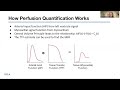
Myocardial blood flow (MBF) can be quantified to detect myocardial ischemia and aid in the diagnosis of coronary artery disease. Myocardial perfusion imaging with cardiac magnetic resonance (CMR) is a current non-invasive, radiation-free imaging method to visualize and measure MBF. There are multiple deconvolution methods to perform this measurement, but no standardized, accessible workflow. The goal of this project was to develop a Python-based computational tool to implement multiple models for deconvolution quantification and allow for systematic comparison and reproducible analysis. The tool processes DICOM images to extract curves of signal intensity over time in the left ventricle and myocardium and implements pre-processing features such as automatic dual-bolus scaling for arterial input function (AIF) estimation. It performs myocardial signal deconvolution to identify a tissue transfer function (TTF) according to either the Fermi model, the Fermi model with a delay parameter, or a “model-free” transfer function. It automatically calculates MBF from the TTF and produces visualizations of model fits and residuals to assess goodness-of-fit. Simulated testing results demonstrate the ability to produce consistent MBF results validated across model types. This work supports reproducibility and flexibility in perfusion analysis for research and applications in cardiovascular imaging.
Presentation 3
EMMA CORDEIRO, Paul Hirose, and Kymora Scotland
Escherichia coli and Pseudomonas aeruginosa are bacteria which produce biofilm and have been found within calcium oxalate stone samples. We have shown that biofilm leads to the formation of these stones through binding with crystals and facilitating aggregation. A key component of this process is supersaturation of the urine. Urinary supersaturation facilitates calcium oxalate nucleation when nuclei bind and aggregate to bacteria within biofilm. This project aims to identify the role of bacteria and biofilm in altering calcium concentrations and the supersaturation of calcium in the urine. Inductively Coupled Plasma Mass Spectrometry (ICP-MS) will be used to measure calcium concentrations following the inoculation of bacteria into patient urine samples. We hypothesize that local areas of biofilm will have increased calcium accumulation due to its ability to trap calcium ions. Results obtained using urine samples of both kidney stone and non-kidney stone patients will provide insight into how differing urinary conditions play a role in kidney stone pathogenesis. Furthermore, experiments utilizing bacterial inoculation in conjunction with renal epithelial cells can provide substantial insight into how calcium concentrations are modulated in the kidney. This may prove useful in the development of more targeted treatments and for the prevention of calcium oxalate stone development in patients.
Presentation 4
SERENA KIM, Hyae Ran Byun, Seokjo Kang, Terrence Lin, and Warren Tourtellotte
Microglia (MG) are resident phagocytes of the central nervous system (CNS) that maintain brain homeostasis by clearing cellular debris and pathogenic aggregates, including amyloid plaques in Alzheimer’s Disease (AD). In AD mouse models, increased angiotensin-converting enzyme (ACE) expression in mouse MG enhanced their immune function to improve phagocytic activity, neuroprotection, and reduced synapse loss. This project investigates whether human MG into immunodeficient mouse brains could serve as a pre-clinical model for studying human MG function in vivo. Hematopoietic progenitor cells (HPCs) were transplanted into 3–4-week-old immunodeficient MRG mice via stereotaxic brain injections. To facilitate engraftment, endogenous mouse MG were depleted using the CSF1R inhibitor PLX-3397. Immunofluorescence (IF) staining of brain sections was performed using antibodies to Iba1 to label all MG and Ku80 was used to specifically identify human-derived cells. Results demonstrated robust engraftment, with approximately 80% of mouse brain MG populations replaced, and over 99% of Iba1+ cells identified as human MG. Preliminary findings indicate that these grafts remain stable for at least one-year post-transplantation. This in vivo model system makes it possible to study the function of modified human MG as a powerful platform to study human-specific neuroimmune mechanisms. Modified human MG may be useful in transplantation therapy for treating neurodegenerative diseases such as Alzheimer’s Disease.
Presentation 5
SOFIA KRIVITSKY, DYLAN OEST, KIERA SHETH, ARIV TANDON, Gabriel Munoz, Lewis Simon, Anjay Rastogi
IgA Nephropathy (IgAN) is characterized by the pathological deposition of immunoglobulin A (IgA) in the glomeruli, leading to chronic inflammation and progressive impairment of renal filtration. Symptoms of IgAN include excessive blood (hematuria) and protein in the urine (proteinuria), with possible manifestation of end-stage kidney disease through progressive scarring. Investigational therapies could prove to be effective in slowing or halting the progression of IgAN. This study aims to evaluate the efficacy, safety, and pharmacokinetics of sefaxersen in participants with primary IgAN who are at risk of progressive kidney disease. Sefaxersen is an antisense oligonucleotide that inhibits complement factor B, which is a critical focus in disease management for IgAN. This study is a Phase III, randomized, quadruple-blind trial. The main criteria include a biopsy-confirmed IgAN diagnosis, treatment with ACE inhibitors or ARBs for at least 90 days prior, a urine protein-to-creatinine ratio (UPCR) ≥ 1 (can include units here if you want for clarity), and an estimated glomerular filtration rate (eGFR) ≥ 20. Participants are randomized to receive Sefaxersen or a placebo injection, administered at days 1, 15, and 29, then every 4 weeks until week 105. Primarily, participants’ UPCR will be assessed relative to baseline following treatment. Secondary outcomes include changes in eGFR, time to the composite kidney failure endpoint, plasma sefaxersen concentration, treatment-emergent adverse events, symptoms, and quality of life.
Presentation 6
AUBRIENNE SILVA, Steven S. Raman, Zahra Mohammadigoldar, Pedram
Keshavarz, Brian Nguyen, Aarib Shahab, Vatche G. Agopian, Jihane
Benhammou, Richard S. Finn, Justin McWilliams, Jason Chiang,David S. Lu
Purpose: Assess impact of pre-locoregional therapy (Pre-LRT) percutaneous needle biopsy-acquired histopathologic tumor differentiation (PCB-TD) and treatment type on overall survival (OS) in hepatocellular carcinoma (HCC).
Methods: IRB-approved, HIPAA-compliant study. 488 HCC patients (mean age: 64.9) who underwent 18G PCB within 6 months prior to LRT were included. Patients were categorized into well-differentiated (well TD) ( n=153) and moderately/poorly differentiated (mod/poor TD) (n = 335) subcohorts. Treatments included LRT alone (n=57) and LRT followed by orthotopic liver transplantation (OLT) (n=156). OS evaluated using Kaplan-Meier analysis, with the Gehan generalized Wilcoxon test.
Results: 5-year OS was 80.6% for well TD vs 71.8% for mod/poor TD (p=0.001). By treatment, 5-year OS was 89.8% for LRT+OLT and 68.5% for LRT alone. In mod/poor TD, OLT significantly improved OS vs LRT alone (HR 0.31, 95% CI: 0.18–0.51; p=0.001), while no significant difference was observed in well TD (HR 0.61, 95% CI: 0.27–1.35; p=0.228). Kaplan-Meier curves showed clear separation in mod/poor TD and minimal separation in well TD.
Conclusion: Pre-LRT PCB-TD stratifies survival benefits for OLT. Mod/poor TD derive substantial OS benefit from OLT, while well TD show no meaningful survival advantage.
Significance: Pre-treatment biopsy-based tumor differentiation enables risk-adapted treatment selection, supporting prioritization of OLT for patients with aggressive tumor biology.
Presentation 7
CLAIRE SPANO, Shriya Reddy, Caitlin Tang, Alejandro Espinoza, Clove Taylor, Andreas Schwingshackl, Matt Zinter, Daniela Markovic, Matteo Pellegrini, Colin J. Sallee, Anil Sapru
Critically ill patients exhibit distinct inflammatory phenotypes that lead to divergent outcomes and directly impact quality of life. A comprehensive, multi-omic understanding of these subphenotypes is therefore essential for advancing personalized care. Metabolic reprogramming is known to be a driver of immune dysfunction in critical illness, and metabolomic profiling has shown promise in adult patients. However, pediatric applications remain limited, often restricting the generalizability of key findings. To address this severe translational gap, we analyzed both untargeted and targeted metabolomics data derived from plasma samples of a cohort of critically ill pediatric patients. Leveraging longitudinal statistical analyses, we contextualized metabolic features with their associated inflammatory phenotypes. Our preliminary results showed that certain targeted biomarkers were upregulated in severe presentations, demonstrating that clinical heterogeneity across inflammatory phenotypes extends even further, to the metabolomic level. Further analysis has revealed even more complex, unique metabolic signatures of each inflammatory phenotype.These findings provide critical insight into the biological mechanisms underlying pediatric critical illness. By extending metabolomic characterization to pediatric populations, our research contributes to a more precise understanding of disease variability and supports the development of targeted, personalized therapeutic strategies.