Microbiology, Immunology, and Molecular Genetics (MIMG): Prerecorded presentation - Panel 2
Location: Online - Prerecorded
Presentation 1
JULIA BELYSHEVA, Victor Mendoza, Peixiang Zhang, Rebecca Hernandez, Carrie Wiese, Laurent Vergnes, Barbara Soliman, Kelsey Jarret, Thomas Vallim, and Karen Reue
Sex Differences in Postprandial Triglyceride Metabolism are Driven by Androgen-Mediated Hepatic CD36 Suppression
Cardiometabolic diseases - including obesity, type 2 diabetes, and cardiovascular disease (CVD) - are chronic inflammatory conditions associated with the consumption of a high-fat Western-style diet. Males exhibit higher rates of CVD incidence and mortality compared to females, a sex difference that persists even after controlling for body composition differences. Assessing CVD risk typically relies on fasting triglycerides (TG); however, postprandial TG measurements more accurately reflect nutritional physiology and provide a stronger predictor of disease. To determine how biological sex regulates postprandial TG metabolism, we used the Four Core Genotypes mouse model and found that gonadal sex - rather than chromosomal sex - independently governs the postprandial TG response. Mechanistically, testes-derived androgens suppress hepatic CD36 expression, limiting hepatic lipid uptake and thereby sustaining elevated circulating TG levels following a high-fat meal. We validated this mechanism with the sham vs gonadectomized mouse model and are currently conducting in vitro androgen dose-response studies on HepaRG hepatocytes. These findings identify androgen-mediated suppression of CD36 as a key driver of postprandial dyslipidemia and establish a mechanistic basis for sex differences in postprandial lipid metabolism. This work provides a foundation for developing sex-informed therapeutic strategies to improve cardiometabolic disease outcomes.
Presentation 2
KRISTINE CHAI, Katherine Montalvo Reglado, Daniela Enriquez-Ochoa, Wendy F. Liu
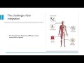
The foreign body response is a major obstacle to biomedical implant integration, involving macrophage fusion into foreign body giant cells (FBGCs) that drive chronic inflammation. While biochemical triggers are well-characterized, the influence of mechanical cues, specifically substrate stiffness, remains poorly understood. Reactive oxygen species (ROS) have emerged as key mediators of this process, functioning as signaling molecules and drivers of oxidative stress. This study uses an in vitro implant-tissue chip model to characterize the effect of stiffness on ROS dynamics and tests the mediation effect of the mechanosensitive ion channel Piezo1.
Bone marrow derived macrophages from mice were seeded on fibronectin coated polyacrylamide coverslips and Mat-Tek dishes, with fusion induced via IL-4 and GM-CSF. To quantify ROS production in response to mechanical cues, live cell analysis was performed using CellROX, MitoSOX, and luminol assays, followed by image analysis. This study evaluates the hypothesis that Piezo1 gain-of-function enhances fusion-associated ROS generation on soft substrates (1 to 10 kPa) to levels comparable with those on rigid substrates (100 kPa).
By characterizing the link between stiffness-induced Piezo1 activation and ROS production, this research clarifies how mechanical microenvironments drive oxidative stress during FBGC formation. Understanding these pathways yields insights for designing targeted interventions to control the foreign body response and improve implant biointegration.
Presentation 3
AAKASH JEYSANKAR, ARUSHI PARIKH, HANA KIM
Monkeypox (Mpox) is an emerging public health concern due to its capacity to sustain human-to-human transmission through close contact. The objective is to investigate the genetic basis of viral spread to be better informed when considering therapeutic developments. This project explores specific regions of the mpox genome which contribute to viral propagation, with the goal of identifying targets for antiviral methods. In order to accomplish this, we have been using our bacterial cloning protocol to design experiments. Our processes include steps such as ligation, transformation, and bacterial inoculation. Initial cloning and screening processes establish a foundation for analyzing gene function through interactions with buffers and compounds using Clonase-based procedures. By analyzing how these genes respond under specific conditions, this approach helps to identify molecular mechanisms of the viral spread. Ultimately, this project establishes a platform via a cloning protocol for the functional characterization of the mpox genome. This baseline contributes to future antiviral developments, and has implications for improving discovery and therapeutic strategies.
COMPASS Scholar
Presentation 4
JAVIER MARTINEZ, Arumugam, Balamurugan, Otto Yang
Engineering Antibody-Pseudotyped Lentiviral Vectors for Targeted Gene Delivery to CD34⁺ and CD123⁺ Hematopoietic Cells
Hematopoietic stem and progenitor cells are essential for immune function and blood formation, developing methods to target CD34- and CD123-expressing cells could enable safer and more effective gene therapies, including in vivo generation of engineered immune cells. Likewise, lentiviral vectors are widely used for gene delivery but are pseudotyped with vesicular stomatitis virus glycoprotein G (VSV-G), which lacks cell specificity and can cause toxicity, limiting targeted therapeutic applications. I aim to investigates whether lentiviral vectors pseudotyped with membrane-anchored anti-CD34 and anti-CD123 antibodies can achieve selective transduction of CD34⁺ and CD123⁺ cells. To establish this system, the heavy and light chains of two anti-CD34 antibodies, My10 and QBEND10, were cloned into separate expression vectors and tested for functional binding using Jurkat-CD34 cells and flow cytometry with appropriate controls. Recombinant QBEND10 demonstrated clear and specific CD34 binding comparable to a commercial antibody, whereas recombinant My10 showed no detectable binding above background, highlighting the importance of experimental validation in antibody engineering. This work identifies QBEND10 as a functional recombinant CD34 antibody and supports the development of antibody-pseudotyped lentiviral vectors for targeted gene delivery, potentially advancing strategies for stem cell research and future in vivo cell-based therapies.
Presentation 6
PHUNG, CALLIE, Sharma, Shipra, Papazian, Sandra, Delgado, Yennifer, Bouhaddou, Mehdi
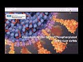
Severe Acute Respiratory Syndrome Coronavirus 2 (SARS-CoV-2) is the virus responsible for the COVID-19 pandemic that claimed the lives of more than 7.1 million individuals around the world. Encoded by SARS-CoV-2 is the accessory protein ORF9b, which has been identified as a key mediator of immune evasion through its interaction with mitochondrial import receptor TOM70. Phosphorylation of Orf9b has been shown to prevent TOM70 interaction, thus enabling downstream interferon responses against SARS-CoV-2 infection. Once phosphorylated, however, the fate of Orf9b has not been determined. This study shows that mutation at S50 and S53 are not only important phosphorylation sites, but also impact localization due to possible implications in proper protein folding. Confocal microscopy shows that phosphodead Orf9b does not mimic the localization of native Orf9b, while phosphomimetic Orf9b exhibited more diffuse puncta but increased localization near the plasma membrane in comparison to phosphodead and control. We hypothesize that phosphorylation of S53 redirects Orf9b from its function in innate immune antagonism to viral assembly. We will employ a viral-like particle (VLP) delivery system to directly quantify assembly of phosphomimetic Orf9b in comparison to phosphodead Orf9b and control. The outcomes of this study will shine light on the potential of a "switch" function of Orf9b between immune antagonism and virion assembly governed by phosphorylation, refining its role as a regulatory hub in SARS-CoV-2 pathogenesis.
Presentation 7
LEILANI PRADIS, Eileen Shiuan, Jorge Salcedo-Sifuentes, Robert Prins
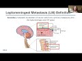
Leptomeningeal Disease (LMD) is a metastatic complication characterized by the dissemination of primary tumor cells into the cerebrospinal fluid (CSF). Despite causing rapid neurological decline and a poor median survival rate of 2 to 7 months, diagnosing LMD remains clinically challenging due to a lack of sensitive standardized tools. The objective of this study was to establish a murine LMD model using LLC-LeptoM lung carcinoma lines engineered with a secreted Gaussia luciferase (sGluc) reporter to enable quantification of tumor burden via liquid biopsy. LLC-LeptoM cells expressing sGluc-mCherry were used to perform in vitro serial dilution assays ranging from 1 to 10^4 cells. For the in vivo model, C57BL/6 mice received injections of 1,000 or 50,000 cells. Tumor burden was quantified by harvesting media or CSF and blood samples, which were then analyzed for bioluminescence after the addition of coelenterazine (CTZ). In vitro validation demonstrated high sensitivity, with bioluminescence detectable from as few as10 cells, exceeding wild-type controls. In vivo, tumor-secreted signal in the CSF was significantly higher than PBS controls in both cell cohorts. This study developed a murine LMD model that enables highly sensitive tracking of tumor burden through liquid biopsy, overcoming limitations of traditional models. By detecting signals in volumes as small as 0.25 µL or 10 cells, this model serves as a validation platform for future LMD research and the development of diagnostics.