Molecular, Cell, and Developmental Biology (MCDB): Prerecorded presentation - Panel 2
Location: Online - Prerecorded
Presentation 1
MALACHI ARP, CAROLYN BREWER, VALERIE JUN, Kashmira Khair, Michelle Yeung, Maitreyi Bharath, Aren Kasparian, Joey Lin, Kelly Tang, Jaunian Chen
This abstract has been withheld from publication.
Presentation 2
ADAM DÉRY, Brian Cheng and Jerzy W. Kupiec-Weglinski
HuR is an RNA-binding protein that regulates the 3' untranslated region (UTR) of target mRNAs involved in ischemia-reperfusion injury (IRI). IRI is a condition caused by hyperactive innate immune signaling leading to liver injury. CEACAM1 (CC1) is a transmembrane glycoprotein that acts as a checkpoint regulator of IR stress. HuR and CC1 mitigate IRI following liver transplantation (LT), though their coordinated role remains undetermined. WT and HuR-KO C57BL6 (8 weeks) were administered LPS/D-Galactosamine (L/D, 400mg/Kg, i.p.) for 6 h before sampling in a model of acute liver injury. Hypoxia and reoxygenation (H/R), Immunofluorescence, and Western Blots were used to evaluate how HuR influences CC1 expression/function. Human LT biopsies obtained before or 2 h after reperfusion were used to determine the incidence of early allograft dysfunction (EAD). Human discard livers aimed to demonstrate the importance of the HuR/CC1 axis. Hepatocyte-specific pre-injury HuR-null mice exhibited more cell death and CC1 expression, reflecting its importance in resolving tissue-specific injury. HuR-targeting short-activating RNAs (saRNAs) preferentially induced the cytoprotective version of CC1 (CC1-S). In the clinical arm, increased HuR and CC1 expression were associated with reduced proinflammatory phenotype and a lower incidence of EAD in patients with LT (n = 164). These findings suggest that HuR's regulation of CC1 is important for the donor tissue quality and offers a potential therapeutic target for future strategies in LT recipients.
Presentation 3
PRADNYA R. KADAM, Nikta Seyedin, Andrea Eastes, Cris Beninca, Orian Shirihai
Type 2 Diabetes (T2D) is a disease characterized by dysfunction in a set of pancreatic endocrine islet cells, known as beta cells, causing insulin resistance and sugar buildup within the body. Mechanistically, beta cells rely on mitochondria, the powerhouse of the cell, as molecular sensors that detect glucose and produce ATP, leading to insulin secretion. However, under conditions of oxidative stress, the beta cells and mitochondria become overworked, damaging the mitochondria and impairing the insulin secretion process. Though current T2D treatments focus on increasing insulin secretion or glucose excretion, mitochondria merit focused investigation. While T2D is extensively researched, there are many limitations with studying beta cells like the fact that animal cell models typically cannot recapitulate human beta cell function, and that human beta cells can only be obtained after death of a patient, making them valuable to use for research. This underscores the importance of using publicly available human islet data to conduct investigations. Here, we aim to link mitochondrial dysfunction to the T2D disease state, using data from the Human Islet Project, a publicly available islet database, by correlating mitochondrial genes to donor characteristics. Our results demonstrate that there is a down-regulation in mitochondrial dynamics proteins like RHOT1, MFN2, and OPA1 for T2D patient populations, alluding to impairments in mitochondrial quality control processes.
Presentation 4
SUMMER A. KELSO and Pei Yun Lee
The following experiments tested whether the purple sea urchin (Strongylocentrotus purpuratus) gene netrin-1-like could be identified, amplified, cloned, and validated within a pGEM-T Easy plasmid vector. Netrin-1-like, a chemotropic axon guidance molecule involved in the developing nervous system, was identified through BLASTn and BLASTx analyses and further supported by domain analysis using InterPro. Phylogenetic analysis was consistent with this identification, demonstrating that purple sea urchin netrin-1-like groups with netrin-1 homologs across multiple species. Gene-specific primers were used to amplify the sequence via PCR, followed by silica column purification and ligation into the pGEM-T Easy vector. The ligation products were transformed into competent Escherichia coli cells and successful integration was selected for using blue-white screening. EcoRI restriction digest and visualization using gel electrophoresis demonstrated the presence of the correct gene insert. While poor DNA quality limited complete validation by Sanger sequencing, alignment of high-quality read segments supported successful cloning. These results indicate successful cloning of the purple sea urchin gene netrin-1-like and establish a foundation for investigating its role in sea urchin development and conserved axon guidance pathways, providing insight into nervous system development.
Presentation 5
NICK J. MARTIN and Pei Yun Lee
The genome of the sea urchin Strongylocentrotus purpuratus has been sequenced, but the specific function of each gene has not been studied. Considering this species’ extensive use as a model organism, it is beneficial to analyze its gene products and the effects they may induce in the organism. A UDP-glucuronsyltransferase (UGT) gene was identified using a BLAST genome database search, with verification of this result using Echinobase and InterPro. A phylogenetic tree was developed to verify gene identity by comparing the coding region to similar genes in other species. Following gene identification, the sequence could then be cloned into a plasmid vector. Utilizing PCR with gene specific primers to amplify the gene allowed it to be ligated into a pGEM-T plasmid vector, which could then be inserted into competent bacteria cells via transformation. Verification of the successful insertion of this gene into the vector was visualized using a restriction digest and sequencing the plasmid. The verification techniques demonstrated the success of the cloning procedure. Further study on the specific protein product of this gene can now be conducted, possibly starting with its function or localization within the sea urchin.
Presentation 6
TALIA PERELLI-MINETTI and Pei Yun Lee
Histone-lysine methyltransferase 2C is a DNA binding enzyme involved in early development and gene regulation. Defects in the protein’s function are associated with cancer and developmental disorders. Using the model organism of S. pupuratus, the histone-lysine methyltransferase sequence was analyzed for functional domains, conservation across species, and sequence variation using Echinobase, Interpro, and NCBI databases along with phylogenetic tree generation and multisequence analysis. The results found multiple DNA binding domains, small-molecule binding domains, and the expected animal homology. Methods for molecular cloning included DNA isolation and amplification, bacterial transformation, plasmid isolation, and sanger sequencing. Molecular cloning resulted in 11 successful transformations, where two of the three selected colonies contained the histone methyltransferase 2C. These transformations were confirmed using plasmid PCR and restriction digest assays. From the results, we conclude that the histone-lysine methyltransferase 2C has functional domains that align with methyl-donating function and argue that the protein sequence is highly conserved across phyla. The high conservation of functional domains, particularly domains for DNA interaction, allows for the conclusion of protein necessity in development. Future directives can investigate where and when the methyltransferase is needed and how disruption of this function causes disease.
Presentation 7
ALICIA SEPULVEDA, Aaron M. Ambrus, David Jelinek, and Hilary A. Coller
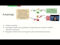
Chronic wounds are medical conditions faced by over 10 million people in the United States alone, characterized by delayed and irregular wound healing. Typically, wound healing proceeds through balanced phases of inflammation, proliferation, and maturation. Chronic wounds have been associated with an upregulated inflammatory response, pinpointing this as an interesting area for studying the potential causes of the condition. Autophagy, a critical homeostatic process that principally recycles, degrades, and repurposes intracellular molecules, is largely involved in cellular proliferation. We studied the role of autophagy in promoting wound repair and regulating inflammation and found that mice with genetic deficiencies in common autophagy genes such as Atg7 and Atg16L1 replicate the delayed healing phenotype of chronic wounds. Additionally, we studied how an inhibition of the canonical NF-kB inflammatory pathway using the small molecule inhibitor, JSH-23, affects wound healing in mice with autophagy deficiency localized to Fsp1-expressing cell types. We found that NF-kB inhibition partially rescues the delayed-healing phenotype. A more direct genetic knockout of the NF-kB pathway was studied in autophagy proficient mice, characteristically improving wound healing. This creates promising results for a rescue phenotype in future experiments that will combine autophagy deficiency with canonical NF-kB inhibition, and highlights the induction of autophagy as an avenue of treatment for chronic wounds.
Presentation 8
EVA WONG, Ivan A. Lopez
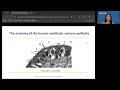
Loss of inner ear hair cells (HCs) and supporting cells (SCs) from aging, noise, ototoxic drugs, or infection causes permanent hearing and balance deficits. Mammalian HCs and SCs lack postnatal regenerative capacity. In mice, deletion of the Cyclin Dependent Kinase Inhibitor 1B (CDKN1B, p27) enables SC proliferation and limited HC regeneration. This study investigated whether p27 protein expression in the adult human inner ear mirrors the pattern observed in mice. Human temporal bone specimens were obtained post-mortem from 6 subjects documented with a lack of auditory or vestibular symptoms. Fresh utricular maculae and cristae ampullaris tissues were surgically obtained from 5 patients with acoustic neuromas included. Cryostat tissue sections (20 microns thick) were incubated with primary antibodies against p27 (1:100, rabbit polyclonal antibody), followed by HRP-DAB-tagged secondary antibodies. Imaging was performed using an Olympus BX51 fluorescent microscope with an Olympus DP70 digital camera. p27 localized to nuclei of SCs of the cochlear organ of Corti, but not in inner/outer HCs, spiral ligament, or stria vascularis. In the human macula utricle and cristae ampullaris sensory epithelium, p27 was uniformly expressed in SC nuclei along the basal membrane. Immunocytochemical detection of p27 in cell nuclei was consistent and reliable. The human inner ear p27 expression pattern agrees with published reports of newborn and adult mice inner ear p27 patterns, suggesting it is a valuable hearing regeneration target.